
How Bone Density Testing Works After a Fracture
- T. Armstrong

- 8 hours ago
- 9 min read
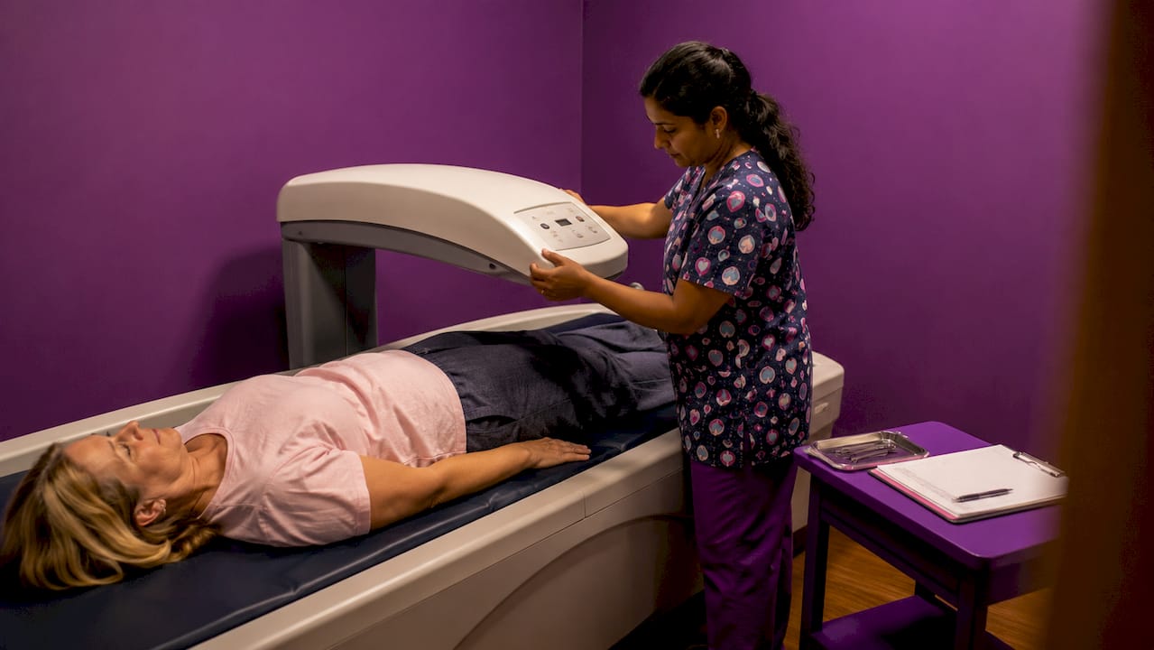
Bone density testing measures the mineral content in your bones to estimate how likely you are to fracture again — not to diagnose the break you already have. That distinction matters for your recovery. According to NIH/NIAMS, the test identifies osteoporosis and predicts future fracture risk, giving your care team a baseline to guide treatment decisions. The MedlinePlus clinical encyclopedia confirms that radiation exposure from a DXA scan is extremely low — lower than a standard chest X-ray — so safety is rarely a barrier.
If you’ve recently had a fracture, here’s why this test belongs on your radar:
Guides treatment: Results help your clinician decide whether medication, supplements, or closer monitoring is the right path.
Establishes a baseline: Your first scan becomes the reference point for every future scan.
Supports fall prevention: Low bone density often travels with other risk factors your care team can address.
Fracture-club and the Bone Health & Osteoporosis Foundation (BHOF) both recognize bone health assessment as a core part of fracture recovery — not an afterthought.
Table of Contents
What does bone density testing actually measure?
Bone mineral density (BMD) is the amount of calcium and other minerals packed into a segment of bone, expressed in grams per square centimeter. A higher BMD generally means stronger bone; a lower BMD means higher fracture risk. The NIAMS frames the test as a preventive assessment: it predicts what might break next, not what already broke.
Your DXA report translates your raw BMD into two standard scores:
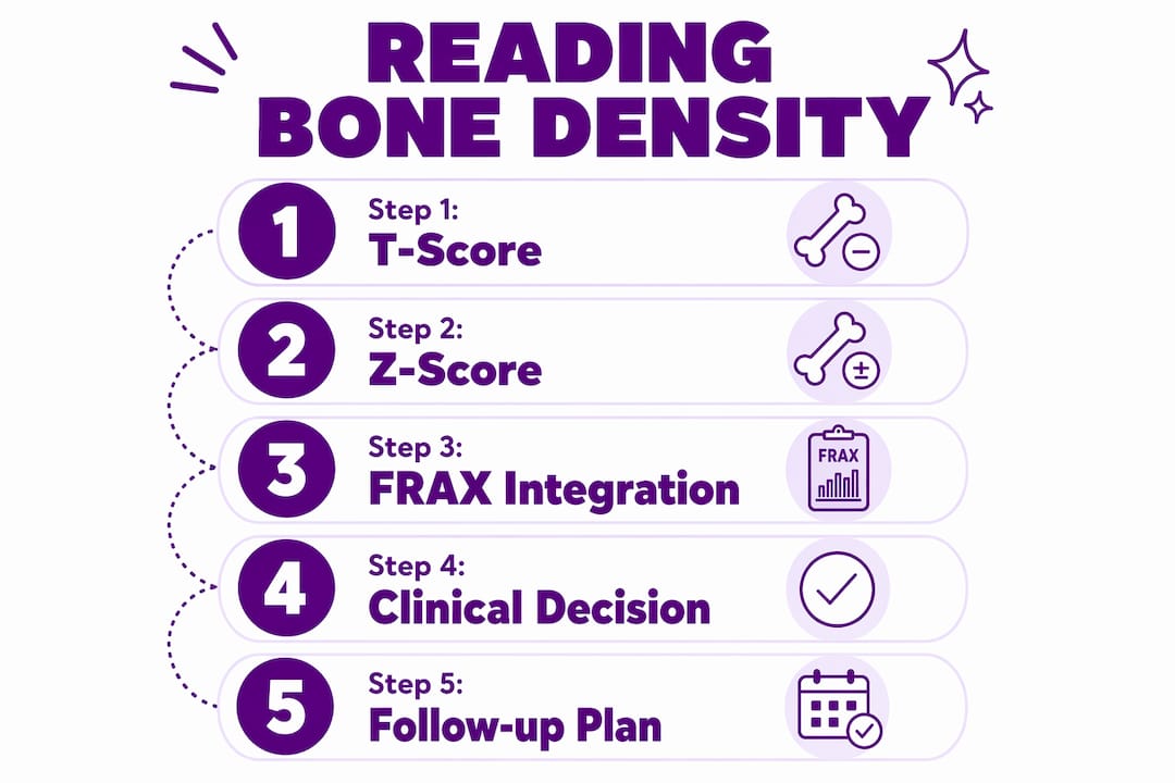
T-score: Compares your BMD to a healthy young adult. This is the primary number used to classify osteoporosis and osteopenia.
Z-score: Compares your BMD to people your own age and sex. Clinicians lean on this score for younger patients or when a secondary cause (medication, disease) is suspected.
Clinicians rarely act on scores alone. Tools like FRAX combine your T-score with age, weight, prior fracture history, and other clinical factors to estimate your 10-year fracture probability. For reading bone health lab results in context, that combined picture is what actually drives treatment decisions.
What types of bone density tests are available?
Test | Site | Best Use | Limitation |
Central DXA | Hip and lumbar spine | Gold standard for diagnosis and monitoring | Requires imaging facility |
Peripheral DEXA (p-DEXA) | Finger, wrist, or heel | Community screening | Less definitive; positive result needs central DXA |
Quantitative CT (QCT) | Spine or hip | Complex cases with hardware or degeneration | Higher radiation; less widely available |
Heel ultrasound | Heel | Radiation-free screening | Lower precision; not used for diagnosis |
DXA (dual-energy X-ray absorptiometry) is the clinical standard for diagnosing osteoporosis and monitoring treatment response. It scans the hip and lumbar spine — the sites most likely to fracture in people with low bone density.
Peripheral DEXA devices are portable and show up at health fairs or pharmacies, but a low result there is a prompt to get a central DXA, not a final diagnosis. Quantitative CT can separate trabecular from cortical bone and is sometimes ordered when spinal hardware or severe degeneration makes DXA readings unreliable.
One important distinction: a bone scan is not the same as a bone density test. A bone scan uses an injected radioactive tracer to detect active problems like infection, cancer, or an acute fracture. DXA involves no injection and measures mineral density only.
Pro Tip: If a peripheral screening test shows low density, ask your clinician for a central DXA before making any treatment decisions. Peripheral results are a flag, not a verdict.
What should you expect at a DXA appointment?
A DXA scan is a quick, painless X-ray test that uses two low-energy X-ray beams to measure bone mineral content. Most appointments run a short duration from start to finish.
Before you go:
Wear loose clothing without metal zippers, buttons, or underwire.
Skip calcium supplements for 24 hours before the scan if your clinician advises it.
Tell the scheduling team if you are pregnant — DXA is typically avoided during pregnancy.
During the scan: You lie on a padded table while the scanner arm passes slowly over your hip and lower spine. Technologists use foam blocks and positioning aids to rotate the hip inward for accurate measurements. If you have a cast, limited range of motion, or surgical hardware, let the technologist know before the scan begins — they can adjust positioning or document the limitation in your report.
You hold still for a few seconds at a time while each image is captured. No needles, no injections, and radiation exposure is extremely low — lower than a chest X-ray.
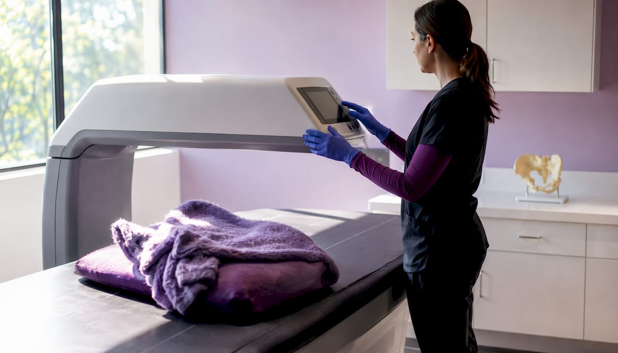
Pro Tip: Mention any recent imaging, metal implants, or casts to the technologist before the scan starts. Hardware near the scan site can skew your T-score, and a note in the report protects the accuracy of future comparisons.
For a full step-by-step walkthrough, the bone density test preparation guide covers everything from what to eat to what to bring.
How do you read your results, and where does FRAX come in?
Your DXA report gives you a T-score and a Z-score. The T-score is the one most clinicians focus on for adults over 50.
T-score range | Classification | What it typically means |
Above −1.0 | Normal | Bone density within expected range |
−1.0 to −2.5 | Osteopenia | Below average; monitor and address risk factors |
−2.5 or lower | Osteoporosis | Significantly reduced density; treatment discussion warranted |
T-score thresholds guide diagnosis and treatment decisions across clinical practice. A T-score between −1.0 and −2.5 doesn’t automatically mean medication — it means your clinician will weigh the full picture.
That’s where FRAX enters. FRAX integrates your femoral neck BMD with age, sex, body weight, prior fracture, glucocorticoid use, smoking status, and family history to calculate a 10-year probability of a major osteoporotic fracture or hip fracture. A T-score of −2.0 in a 50-year-old with no other risk factors looks very different from the same score in a 70-year-old who recently fell. FRAX makes that difference visible.
Z-scores matter most for younger patients or when a secondary cause is suspected. A Z-score well below the expected range prompts investigation into medications, hormonal conditions, or nutritional deficiencies before any bone-specific treatment begins.
Who should get tested, and when?
A recent low-trauma fracture is itself a strong clinical reason to test. Clinical guidance emphasizes that a fragility fracture signals elevated future risk, making testing a logical next step for secondary prevention.
Common triggers for bone density testing in the U.S.:
Any adult with a low-trauma (fragility) fracture
Women 65 and older (USPSTF recommendation)
Younger postmenopausal women with elevated risk factors
Long-term glucocorticoid (steroid) use
Low body weight, family history of osteoporosis, or secondary conditions affecting bone
The USPSTF screening guidance and clinical societies like the BHOF recommend testing when risk is elevated, not as a universal screen for all adults. If you’ve just had a fracture, coordinate the timing of your DXA with your orthopedist or primary care provider — some clinicians prefer to wait until acute healing is underway before establishing a baseline. For a deeper look at what factors drive risk, the types of fracture risk factors guide is a practical starting point.
What bone density testing cannot tell you
BMD testing measures mineral content. It does not measure bone quality, balance, muscle strength, fall risk, or whether an acute fracture is currently healing. Those gaps matter for recovery planning.
Specific limitations to keep in mind:
Acute fractures: DXA cannot detect an active fracture. X-ray, CT, or MRI is used for that.
Hardware interference: Plates, screws, or joint replacements near the scan site can artificially inflate your T-score.
Peripheral tests: Results from wrist or heel devices are not interchangeable with central DXA scores.
Short-term changes: A single scan won’t show week-to-week healing progress.
Pro Tip: Ask your clinician how your BMD results will be used alongside functional assessments, physical therapy goals, and fall-prevention planning. The number is one input, not the whole answer.
Practical next steps after you get your results
Review your results with the clinician managing your fracture care — your orthopedist, endocrinologist, or primary care provider. Then work through this checklist:
Medication: If your T-score and FRAX results suggest treatment is warranted, discuss options with your doctor.
Calcium and vitamin D: Get your levels checked. Deficiencies are common and correctable. The bone health supplements checklist covers dosing considerations.
Nutrition: Protein supports bone healing. The role of protein in bone healing explains why it matters during recovery.
Strength and balance: Ask for a physical therapy referral focused on fall prevention and functional strength.
Home modifications: Remove trip hazards, add grab bars, and improve lighting — small changes that reduce fall risk meaningfully.
Adaptive clothing and dressing aids: If a cast or brace makes dressing difficult, Fracture-club’s recovery wear is designed for exactly this stage.
Pro Tip: Keep a copy of your DXA report and always compare future scans to the same sites — hip and lumbar spine. Consistent scan sites give you the most reliable trend data over time.
Key Takeaways
Bone density testing predicts future fracture risk by measuring bone mineral density, and combining those results with FRAX and clinical factors is what actually guides treatment after a fracture.
Point | Details |
What the test measures | BMD in grams per square centimeter, translated into T-scores and Z-scores for clinical use. |
DXA is the gold standard | Central DXA of the hip and spine is the most accurate and widely used method for diagnosis and monitoring. |
FRAX adds clinical context | FRAX combines your T-score with age, prior fracture, and other risk factors to estimate 10-year fracture probability. |
A fracture triggers testing | Any low-trauma fracture is a strong clinical reason to get a bone density test and establish a treatment baseline. |
Fracture-club supports recovery | Fracture-club offers adaptive recovery wear for easier dressing during immobilization, with a portion of proceeds benefiting the BHOF. |
What recovering from a fracture taught us about bone health
Most people who come to Fracture-club are focused on the immediate problem: how to get dressed, how to stay comfortable, how to feel like themselves again. Bone density testing can feel like a distant clinical concern when you’re managing a cast and a recovery schedule. But here’s the perspective worth holding onto: the test is not about what already happened. It’s about what you can prevent next.
The gap most recovery guides miss is the connection between that DXA number and the daily decisions that actually move the needle — nutrition, movement, fall prevention, and having the right support around you. Understanding your T-score is useful. Acting on it, with a care team and a community behind you, is what changes outcomes.
Fracture-club was built around that idea. Recovery wear that makes dressing manageable, community stories that remind you you’re not alone, and a partnership with the BHOF that puts resources directly in your hands. You can explore the bone health best practices guide for ongoing prevention strategies, or browse the blog for recovery stories and practical tips from people who’ve been exactly where you are.
Recovery wear that makes the hard days a little easier
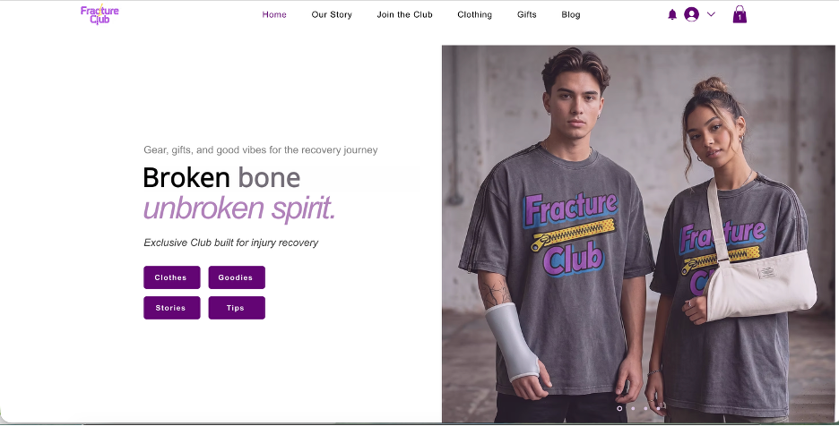
Getting dressed with a cast or brace is genuinely frustrating. Buttons, zippers, and tight waistbands that were never designed for immobilization make a simple task feel like a project. Fracture-club’s adaptive recovery clothing is built for this exact situation.
[
](fracture-club.com)
The adaptive recovery pants feature side magnetic zippers so you can get them on and off without bending or struggling around a lower-limb cast. For upper-body injuries, the easy-on/off sweatshirt opens wide enough to accommodate a sling or shoulder brace without pulling. Both are designed for comfort and function during the weeks when independence matters most. Visit Fracture-club to find the right fit for your recovery stage.
Authoritative sources and further reading
NIH/NIAMS — Bone Mineral Density Tests: What the Numbers Mean: Defines BMD testing, T-scores, and the test’s preventive role.
MedlinePlus — Bone Mineral Density Test: Clinical overview including radiation safety and pregnancy guidance.
MedlinePlus — Bone Density Scan: Plain-language explanation of T-score and Z-score thresholds.
Mayo Clinic — Bone Density Test (DXA/DEXA Scan): Covers test types, peripheral DEXA, and the difference between DXA and a bone scan.
Cleveland Clinic — DXA Scan: Procedure details including positioning and technologist technique.
USPSTF — Osteoporosis Screening: U.S. screening recommendations and FRAX integration guidance.
Bring a printout of any of these sources to your next clinical visit if you want to review specific thresholds or screening criteria with your provider directly.
This article is general health information, not medical advice. Confirm current screening guidelines and treatment options with your clinician or a qualified bone health specialist for your specific situation.
Recommended
Comments